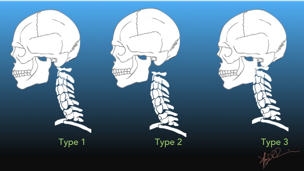

These rare injuries require only external immobilization with an orthosis if there is no associated ligamentous injury. Type I fractures are avulsion fractures of the tip of the odontoid process. Magnetic resonance imaging has been recommended for evaluating associated ligamentous injuries and may be helpful in detecting occult cervical spine fractures. Plain radiography, polytomography, and computed tomography are all useful in delineating the fracture pattern.

Fracture displacement, compromised blood supply, comminution, and iatrogenic distraction have all been implicated in the reported high rates of nonunion. doi: 10.4103/0971-3026.Fractures of the odontoid process are uncommon injuries. CT and MR imaging of odontoid abnormalities: a pictorial review. Jain N, Verma R, Garga UC, Baruah BP, Jain SK, Bhaskar SN.Oblique axis body fracture: an unstable subtype of Anderson type III odontoid fractures-apropos of two Cases. Takai H, Konstantinidis L, Schmal H, Helwig P, Knöller S, Südkamp N, et al.Study on accuracy and interobserver reliability of the assessment of odontoid fracture union using plain radiographs or CT scans. Koller H, Kolb K, Zenner J, Reynolds J, Dvorak M, Acosta F, et al.3 Topics:Ĭomputed tomography, CT, cervical spine, cervical spine fracture, odontoid, orthopedics, spine, neurosurgery. 3 However, if there is concern for ligamentous injury, MRI is the preferred modality. Strict cervical spine precautions must be adhered to until adequate imaging and surgical consultation is obtained.Ĭomputed tomography of the of cervical spine fractures poses several advantages to plain film radiography due to the ability to view the anatomy in three planes. Determining the stability of a Type III Odontoid fracture requires radiographic evaluation. 2 Type 3 odontoid fractures are classified by a fracture of the Odontoid process, as well as the lateral masses of the C2. This fracture is unstable and requires operative stabilization. It is considered a stable fracture. Type 2 is the most common and is a fracture involving the base of the odontoid process, below the transverse component of the cruciform ligament.


Type 1 is the rarest and is a fracture involving the superior segment of the Dens. Odontoid fractures comprise approximately 10% of vertebral fractures, and there are three types with varying stability. Hyperextension or hyperflexion injuries can induce significant stress causing fractures. Unique to C2 is a bony prominence, the Odontoid Process (Dens). The cervical spine is composed of seven vertebrae, with C1 and C2 commonly referred to as the Atlas and Axis, respectively. He was evaluated by orthopedic spine service who recommended conservative, non-operative management. Significant findings:Ĭomputed Tomography (CT) of the cervical spine showed a stable, acute, non-displaced fracture of the odontoid process extending into the body of C2, consistent with a Type III Odontoid Fracture. He was neurovascularly intact, and placed in an Aspen Collar with strict spine precautions. Exam was notable for left parietal scalp laceration and midline cervical spine tenderness with no obvious deformities. An 84-year-old male presented with left-sided posterior head, neck, and back pain after a ground level fall.


 0 kommentar(er)
